The human mouth is a marvel of nature, teeming with intricate structures crucial for our well-being. But lurking within this intricate ecosystem lies a potential invader: the cavity. While the image of a gaping hole in a tooth often comes to mind, early cavities can be surprisingly difficult to detect, presenting a stealthy threat to oral health.
Understanding the Villain: The Stages of Tooth Decay
To effectively identify the early signs of a cavity, it’s essential to understand the stages of tooth decay:
1. Demineralization:
Imagine your enamel, the tooth’s outermost layer, as a fortified shield protecting your inner dental structures. This stage is akin to the villain, harmful bacteria, launching their first attack. These bacteria, fueled by the sugars we consume, produce acidic waste. This acidic environment disrupts the natural mineral balance in your enamel, causing demineralization. This weakening of the enamel is often symptomless and undetected at this stage.
2. Early Cavity (Enamel Caries):
As the acidic assault by bacteria continues, the weakened enamel starts to show signs of vulnerability. This stage is the battleground where early cavity formation, also known as enamel caries, occurs. However, due to the enamel’s inherent transparency, these early signs can be subtle and easily missed:
- Slight Discoloration: You might notice faint white, yellow, or brown spots on the affected tooth surface. These are areas where the enamel has lost some minerals, but the underlying structures remain intact.
- Surface Texture: Run your tongue gently over your teeth. Early cavities might sometimes feel slightly rough or chalky compared to the smooth surface of healthy enamel.
3. Dentin Decay:
If left unchecked, the villainous bacteria breach the weakened enamel and penetrate deeper into the dentin, the softer layer beneath. This stage marks the enemy’s advance and signifies a more critical situation. Here’s why:
- Increased Sensitivity: Dentin contains microscopic tubules that connect directly to the pulp, the tooth’s inner nerve center. As the decay invades the dentin, these tubules become exposed, leading to increased sensitivity to hot, cold, sweet, or acidic foods and beverages.
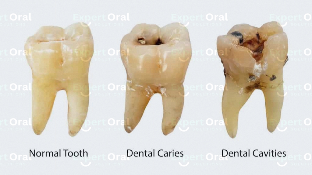
- Visible Damage: Depending on the severity, you might notice a darkening of the discolored area or even the formation of a small cavity on the tooth surface.
4. Advanced Cavity (Pulpal Involvement):
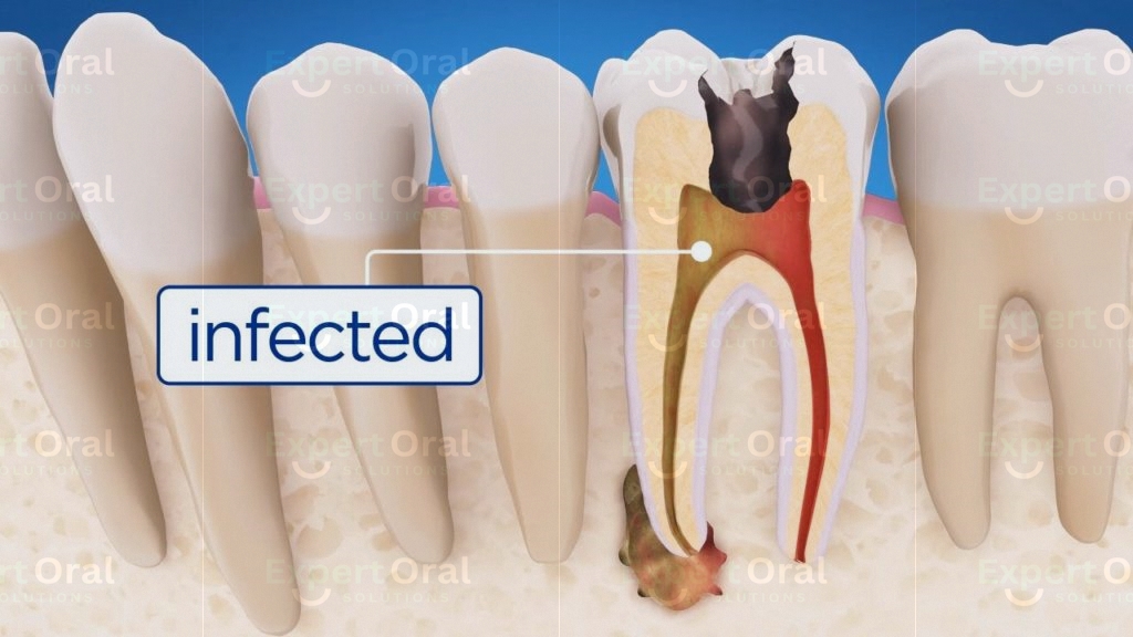
This stage represents the villain’s complete victory if left untreated. The decay now reaches the pulp, the innermost layer of the tooth, containing nerves and blood vessels. This invasion causes significant damage and triggers the following:
- Intense Pain: The exposed pulp becomes highly sensitive and inflamed, leading to throbbing pain that can be spontaneous or triggered by hot/cold stimuli.
- Visible Damage: The cavity becomes significantly larger and deeper, often accompanied by discoloration and potential swelling in the surrounding gum tissue.
The Elusive Early Cavity: What to Look For
Identifying early cavities requires a keen eye and awareness of subtle changes in your teeth. While detection isn’t always straightforward, here are the telltale signs to watch out for:
1. Slight Discoloration:
- Not universally present: Early cavities may not always exhibit noticeable discoloration, making them easily overlooked. However, be on the lookout for faint white, yellow, or brown spots on the tooth surface. These discolored areas indicate demineralization, where the enamel has begun to lose minerals due to bacterial activity.
- Location: These spots often appear on tooth surfaces that come into frequent contact with food and sugars, such as the chewing surfaces (the tops of molars and premolars) or between teeth near the gumline.
2. Surface Texture:
- Run your tongue gently over your teeth, paying close attention to any areas that feel different. Early cavities can sometimes present with a slightly rough or chalky texture compared to the smooth enamel surface of healthy teeth.
- Caution: This method is not foolproof as some healthy teeth might have naturally textured surfaces. Additionally, relying solely on this method could lead to missing cavities in less accessible areas.
3. Pain or Sensitivity:
- Not a reliable indicator: While increased sensitivity to hot, cold, sweet, or acidic foods and beverages can sometimes be a sign of an early cavity, it’s crucial to remember that its absence doesn’t guarantee the absence of a cavity. Early cavities can progress silently without causing any discomfort in the initial stages.
Beyond the Obvious: Additional Considerations
- Location of the cavity: Early cavities are more likely to develop on back teeth (molars and premolars) as these surfaces are harder to clean effectively and have deeper grooves that trap food particles and bacteria.
- Individual variations: The rate of progression and symptoms can vary depending on individual factors like oral hygiene practices, dietary habits, and the composition of your enamel.
Different Types of Dental Cavities
Cavities, also known as dental caries, are a common oral health concern that affect people of all ages. While most people picture a gaping hole in a tooth when they think of a cavity, the reality is that they can come in different forms, each with its own characteristics:
1. Smooth Surface Cavities:
- Location: These cavities develop on the smooth, flat surfaces of teeth, commonly affecting the sides of teeth near the gumline or the chewing surfaces of front teeth.
- Formation: They often arise from plaque buildup due to inadequate brushing and flossing. Plaque harbors bacteria that produce acids, leading to demineralization and ultimately, cavity formation.
- Progression: Smooth surface cavities are relatively slow-growing compared to other types. However, if left untreated, they can eventually progress to affect deeper layers of the tooth.
2. Pit and Fissure Cavities:
- Location: These cavities form in the natural grooves and depressions on the chewing surfaces of molars and premolars. These grooves, called pits and fissures, are often deep and narrow, making them prime targets for plaque buildup and acid formation.
- Formation: Similar to smooth surface cavities, bacteria thrive in these grooves, leading to demineralization and cavity development.
- Progression: Due to the difficulty of effectively cleaning these areas, pit and fissure cavities can progress quickly compared to smooth surface cavities.
3. Root Cavities:
- Location: Unlike the previous two types, these cavities develop on the root surfaces of teeth, the part typically covered by gum tissue.
- Formation: Root cavities are more common in adults, especially those with receding gums. When gums recede, the root surfaces become exposed, making them vulnerable to plaque and acid attack.
- Risk factors: Individuals with gum disease are at a higher risk of developing root cavities due to increased inflammation and plaque accumulation near the gum line.
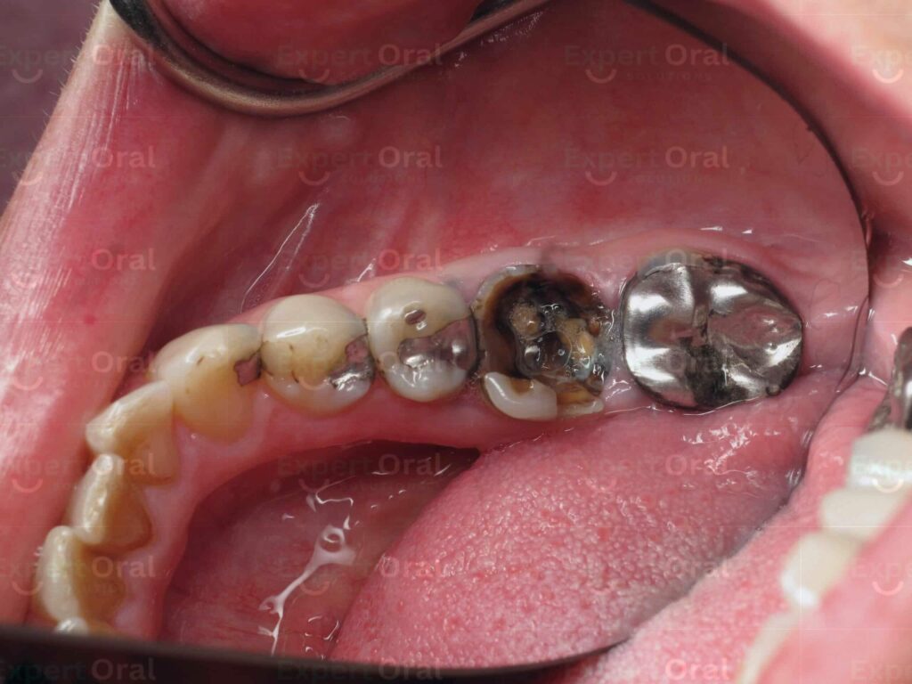
4. Recurrent Cavities:
- Location: These cavities can occur around existing fillings or crowns.
- Formation: They often develop due to inadequate cleaning around restorations, allowing bacteria to thrive and demineralize the surrounding tooth structure.
- Prevention: Maintaining good oral hygiene and regular dental checkups are crucial to prevent recurrent cavities.
Remember: Early detection and treatment of cavities are essential for preserving your oral health and preventing further complications. Regular dental checkups and X-rays allow dentists to identify cavities in their early stages and recommend appropriate treatment options.

Additional Considerations:
- While these are the main types of dental cavities, other less common types exist, such as cervical cavities (affecting the tooth neck) and rampant caries (affecting multiple teeth rapidly).
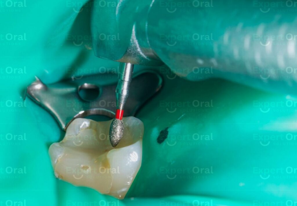
- The severity of a cavity also plays a role in determining its treatment. Early detection allows for more conservative treatments like fluoride applications or fillings, while advanced cavities might require more complex procedures like root canals or extractions.
By understanding the different types of cavities and their characteristics, you can be more vigilant about your oral health and take proactive steps towards prevention.
Differentiating dental cavities from other lesions
Differentiating dental cavities from other lesions can be challenging, especially in their early stages. However, there are some key factors to consider that can help you distinguish between them:
Appearance:
- Cavities:
- Early stages: May show slight white, yellow, or brown discoloration, sometimes appearing rough or chalky to the touch.
- Advanced stages: May exhibit visible holes, cracks, or deeper black or brown discoloration.
- Other lesions:
- Stains: Can appear brown, yellow, or white, but typically have a uniform color and smooth texture. They often don’t change over time unlike cavities.
- Enamel defects: May resemble cavities but usually lack the discoloration or roughness associated with them. They are often present since childhood and don’t change in size or appearance.
Symptoms:
- Cavities:
- Early stages: Might not cause any symptoms, but some individuals experience mild sensitivity to hot, cold, or sweet foods and beverages.
- Advanced stages: Often cause significant pain, sensitivity, and discomfort, especially when biting or chewing.
- Other lesions:
- Stains: Typically don’t cause any pain or sensitivity.
- Enamel defects: Usually asymptomatic.
Location:
- Cavities:
- Smooth surface cavities: Common on flat surfaces of teeth, especially near the gumline or on the chewing surfaces of front teeth.
- Pit and fissure cavities: Occur in the grooves and depressions of chewing surfaces on molars and premolars.
- Root cavities: Develop on the root surfaces of teeth, typically exposed due to receding gums.
- Other lesions:
- Stains: Can appear on any tooth surface, but often occur on front teeth due to increased exposure to staining substances like coffee, tea, or tobacco.
- Enamel defects: Can be present on any tooth surface but are often localized to specific areas.
Progression:
- Cavities:
- Progressive: If left untreated, cavities grow larger and deeper over time, eventually reaching the pulp, causing significant pain and requiring extensive treatment.
- Other lesions:
- Static: Stains and enamel defects generally remain stable in size and appearance over time unless they are caused by ongoing factors like poor oral hygiene or continuous stain-causing habits.
It’s important to remember that these are general guidelines, and accurate diagnosis always requires professional expertise.
Here are some additional points to consider:
- Dental X-rays: Can provide valuable insights into the presence and extent of cavities, especially in their early stages when they might not present visible signs.
- Professional examination: A dentist can perform a visual examination, assess sensitivity, and use specialized tools to differentiate between cavities and other lesions.
- Don’t self-diagnose: Attempting to self-diagnose can lead to delayed treatment and worsen the issue.
Beyond the Naked Eye: Seeking Professional Help
Despite your best efforts, early cavities can be challenging to detect at home. Here’s where your dentist becomes your ultimate ally:
- Regular Dental Checkups: Scheduling regular dental checkups and cleanings every six months is crucial. Dentists possess the expertise and tools, like dental X-rays, to identify cavities at their earliest stages and recommend the appropriate treatment.
- Early Detection, Early Prevention: Catching cavities early allows for minimally invasive and cost-effective interventions like fluoride applications or remineralization treatments.
Authentic Research References:
- American Dental Association (ADA): https://www.ada.org/en/about/press-releases/american-dental-association-releases-new-tooth-decay-treatment-guideline
- National Institute of Dental and Craniofacial Research (NIDCR): https://www.healthline.com/health/dental-and-oral-health/tooth-decay-stages
- Mayo Clinic: https://parkdale-dental.com/ottawa-dental-blog/tooth-decay/how-do-cavities-form-in-your-teeth/
Remember, prevention is always better than cure. Maintaining good oral hygiene practices like brushing twice a day, flossing daily, and limiting sugary foods can significantly reduce your risk of cavities. Additionally, scheduling regular dental checkups allows for early detection and intervention, protecting your smile and overall health in the long run.
So, be vigilant, observe your oral health closely, and consult your dentist regularly. By understanding the early signs of cavities and taking proactive steps, you can maintain a healthy, confident smile for years to come!


